

We addressed bioproduction from a materials physics perspective. One research axis began with physico-chemical studies of nanoparticle biotransformations within cells, which led to a new line of investigation focused on the endogenous production of nanoparticles in eukaryotic cells, analogous to biomineralization processes in prokaryotes.
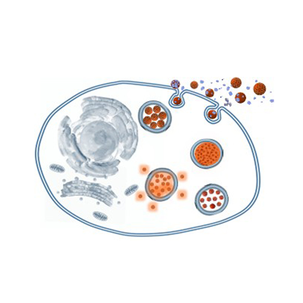
In parallel, we expanded into the bioproduction of extracellular vesicles (exosomes) by our cell systems. We established key principles for their nanoengineering and, importantly, developed high-throughput production methods to enhance both yield and cargo loading of extracellular vesicles.
NANOPARTICLES BIOTRANSFORMATIONS AND BIOPRODUCTION
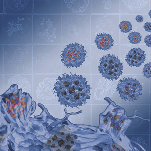
With the growing medical use of magnetic nanoparticles, in particular for cell therapies, it is crucial to study their long-term intracellular fate within living tissues. We provided multiscale quantitative methods to study intracellular iron oxide nanoparticle degradation, showing an unexpected near-total nanoparticle degradation during long-term maturation of a stem cell model tissue. In brief, we developed stem cell spheroids as new biological tools to monitor intracellular nanoparticle degradation, and we managed to perform single spheroid magnetism in situ as a fingerprint of nanoscale transformations. Remarkably, and unexpectedly, the nanoparticles were found to be more than 90% purged inside the tissue in the first ten days of tissue maturation, barely affecting cellular iron homeostasis. The same massive degradation was recapitulated at the single endosome level, using a unique approach based on single purified endosome nano-magnetophoresis.
These results evidencing for the first time a total breakdown of nanoparticles by endosomes in stem cells composing a model tissue bodes well for their safety in medical applications, especially regenerative medicine.
Beyond its obvious nanosafety implications, the impact of the cellular environment on nanomaterials also raises concerns as to their therapeutic applicability, for either the tissue engineering field (long-term stimulation of engineered tissues), or for cancer therapies (serial treatments). It can thus be beneficial to protect iron oxide nanoparticles to massive intracellular dissolution.
We have showed that a gold shell can prevent the intracellular biodegradation of iron oxide nanoflowers and thereby maintain their potential for magnetic hyperthermia, in addition to the excellent photothermal efficiency of the gold shell itself. Besides, it demonstrates that not only magnetic metrics, but also thermal ones can be quantitative mirrors of the intracellular status.
Finally, we were the first to have just evidenced the possible biosynthesis of nanoparticles by human stem cells, using the ions delivered by the degradation of administered chemically synthesized nanoparticles
Selection of publications
- Elucidating the Dynamics of Biodegradation and Biosynthesis of Magnetic Nanoparticles in Human Stem Cells. Curcio A, Curé G, Espinosa A, Menguy N, Galarreta‐Rodriguez I, Abou‐Hassan A, Piquet B, Motte L, Lalatonne Y, Wilhelm C, Van de Walle A. Small 20, 2407034 (2024)
- Biomineralization of magnetic nanoparticles in stem cells. Fromain A, Van de Walle A, Curé G, Péchoux C, Serrano A, Lalatonne Y, Espinosa A, Wilhelm C. Nanoscale 15, 10097-10109 (2023)
- Massive Intracellular Remodeling of CuS Nanomaterials Produces Nontoxic Bioengineered Structures with Preserved Photothermal Potential. A Curcio, A Van de Walle, E Benassai, A Serrano, N Luciani, BB Manshian, A Sargsian, S Soenen, A Espinosa, A Abou-Hassan, C Wilhelm. ACS nano 15, 9782-9795 (2021)
- Transformation Cycle of Magnetosomes in Human Stem Cells: From Degradation to Biosynthesis of Magnetic Nanoparticles Anew. Curcio A, Van de Walle A, Serrano A, Preveral S, Péchoux C, Pignol D, Menguy N, Lefèvre CT, Espinosa A, Wilhelm C. ACS nano 14, 1406-1417 (2020)
- Ever-Evolving Identity of Magnetic Nanoparticles within Human Cells, the Interplay of Endosomal Confinement, Degradation, Storage, and Neo-Crystallization. Van de Walle A, Kolosnjaj-Tabi J, Lalatonne Y, Wilhelm C. Accounts of chemical research 53, 2212-2224 (2020)
- Real-time in situ magnetic measurement of the intracellular biodegradation of iron oxide nanoparticles in a stem cell-spheroid tissue model. Van de Walle A, Fromain A, Plan Sangnier A, Curcio A, Lenglet L, Motte L, Lalatonne Y, Wilhelm C. Nano Research 13, 467-476 (2020)
- Biosynthesis of magnetic nanoparticles from nano-degradation products revealed in human stem cells. Van de Walle A, Plan Sangnier A, Abou-Hassan A, Curcio A, Hémadi M, Menguy N, Lalatonne Y, Luciani N, Wilhelm C. PNAS 116, 4044-4053 (2019) https://www.cnrs.fr/en/what-happens-magnetic-nanoparticles-once-cells
- Impact of magnetic nanoparticle surface coating on their long-term intracellular biodegradation in stem cells. Plan Sangnier A, Van de Walle A, Le Borgne R, Motte L, Lalatonne Y, Wilhelm C. Nanoscale, 11, 16488 (2019)
- Intracellular Biodegradation of Ag Nanoparticles, Storage in Ferritin, and Protection by Au Shell for Enhanced Photothermal Therapy. Espinosa A, Curcio A, Cabana S, Radtke G, Bugnet M, Kolosnjaj-Tabi J, Péchoux C, Alvarez-Lorenzo C, Botton GA, Silva AKA, Abou-Hassan A, Wilhelm C. ACS nano 12, 6523–6535 (2018)
- Magneto-Thermal Metrics Can Mirror the Long-Term Intracellular Fate of Magneto-Plasmonic Nanohybrids and Reveal the Remarkable Shielding Effect of Gold. Mazuel F, Espinosa A, Radtke G, Bugnet M, Neveu S, Lalatonne Y, Botton GA, Abou-Hassan A, Wilhelm C. Advanced Functional Materials, 27, 1605997 (2017)
- Massive Intracellular Biodegradation of Iron Oxide Nanoparticles Evidenced Magnetically at Single Endosome and Tissue Levels. Mazuel F, Espinosa A, Luciani N, Reffay M, Le Borgne R, Motte L, Desboeufs K, Michel A, Pellegrino T, Lalatonne Y, Wilhelm C. ACS Nano, 10, 7627- 38 (2016)
EXTRACELLULAR VESICLES NANO-ENGINEERING
To deliver nanoparticles on-site, the last decades have seen the development of a variety of drug delivery systems. Recently, membrane-bound EVs released by cells, have emerged as a cell-to-cell communication system which could be used to bio-camouflage drugs.
We pioneered this possibility to load EVs with NPs and drugs, by incorporating them in the parent cells prior to trigger vesicles emission. To make such bio-camouflaged NPs a reality for clinical applications, the community must solve production and low loading issues. We showed for instance that an hydrodynamic stimulation can increase the number of EVs (10 times more in 10 times less time), as well as the loading of a drug, and that a laser stimulation (activating a photosensitizer) can also increase EVs production.
Selection of publications
- Massive release of extracellular vesicles from cancer cells after photodynamic treatment or chemotherapy. Aubertin K, Silva AKA, Luciani N, Espinosa A, Djemat A, Charue D, Gallet F, Blanc-Brude O, Wilhelm C. Scientific Reports, 6, 35376 (2016)
- Combining magnetic nanoparticles with cell derived microvesicles for drug loading and targeting. Silva AKA, Luciani N, Gazeau F, Aubertin K, Bonneau S, Chauvierre C, Letourneur D, Wilhelm C. Nanomedicine NBM, 11, 645–655 (2015)
- Cell-derived Vesicles as a Bioplatform for the Encapsulation of Theranostic Nanomaterials. Silva AKA, Di Corato R, Luciani N, Chat S, Gazeau F, Pellegrino T, Wilhelm C. Nanoscale. 7, 11374-84 (2013)
- Magnetic and photoresponsive theranosomes: translating cell-released vesicles into smart nanovectors for cancer therapy. Andriola A, Kolosnjaj J, Bonneau S, Marangon I , Bogetto N, Aubertin K, Clément O, Bureau MF, Luciani N, Gazeau F, Wilhelm C. ACS nano 7, 4954–4966 (2013)
- Mesenchymal stem cell derived extracellular vesicles loaded with at least one photosensitizer and uses thereof for the treatment of peritoneal carcinomatosis Pocard M, Gazeau F, Marangon I, Pinto A, Silva A, Piffoux M, Wilhelm C. Patent Number: EP4061388A1, Publication Date: 2022-09-28, Application Number: EP20807775.0 (2022)
EXTRACELLULAR VESICLE HIGH THROUGPUT BIOMANUFACTURING
Since the beginning of the century, extracellular vesicles (EVs), including exosomes, have emerged as key biological entities whose importance has rapidly expanded over the past decade. These fully biological nanoparticles are released by cells into their environment and play central roles in intercellular communication, regulating a wide range of physiological processes. They transfer proteins, lipids, nucleic acids, and sometimes oncogenic signals, thereby contributing to organ function and systemic regulation.
A key challenge for clinical translation is their large-scale production. We first patented the use of commercial bioreactors for high-throughput EV production by increasing hydrodynamic stresses to induce turbulent regimes. However, this repurposed approach is not optimized for EV production, and dedicated systems are still lacking (EP 2330543131.1, filed 28/03/2023).
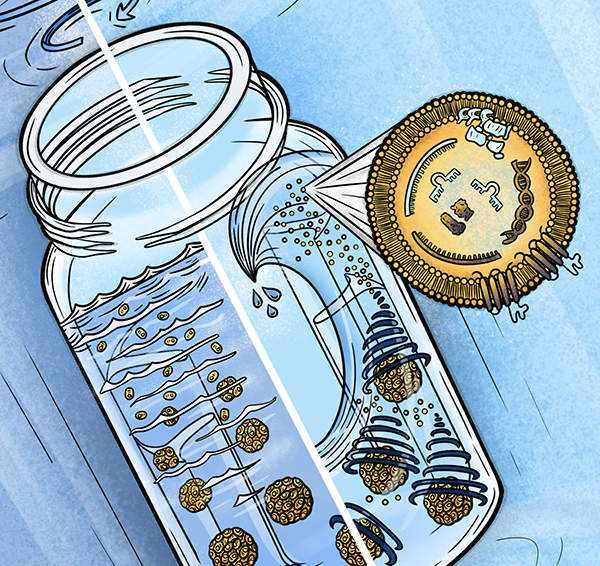
To address this, we developed a modular rotating-tube platform with adjustable hydrodynamic conditions (rotation speed, inversion frequency, and volume). This system enables a fully integrated workflow in which stem cells are first matured into spheroids and then directly used as EV-producing units under higher shear conditions. This biomimetic 3D configuration enhances EV yield and induces specific proteomic signatures associated with improved angiogenic, wound-healing, and anti-inflammatory properties.
In parallel, we developed a cross-slot microfluidic device generating a stable vortex that traps spheroids and significantly enhances EV production (EP24305327.9, filed 04/03/2024).
This technology also extends to bacterial EVs, a field still in its early stages despite strong therapeutic potential, including vaccine development and applications in cancer and metabolic diseases. Using our system, we achieved 10- to 100-fold increases in EV yield compared to standard conditions for both Gram-negative (E. coli) and Gram-positive (Bacillus subtilis) bacteria (EP 24306343.5, filed 08/08/2024).
Finally, we demonstrated that this platform can also be used for the production of lentiviruses and virus-like particles (VLPs), with direct applications in CAR-T cell manufacturing for immunotherapy.
Selection of publications
- Hydrodynamic Bioreactor for High-Yield Production of Extracellular Vesicles from Stem Cell Spheroids with Defined Cargo Profiling. Lenoir S, Thouvenot E, Gropplero G, Dec L, Loew D, Théry C, Perez JE, Wilhelm C. Advanced Science 13, e10607 (2025)
- High-yield bioproduction of extracellular vesicles from stem cell spheroids via millifluidic vortex transport. Thouvenot E, Charnay L, Burshtein N, Guigner J-M, Dec L, Loew D, Silva AKA, Lindner A, Wilhelm C. Advanced Materials, 2412498 (2025)
- Method for efficient production of biological particles comprising a viral or virus-like component in baffled rotating vessel. European Patent Application number EP24306529.9, filed sept 17th 2024. B Lapin, ZL Gouveia, F Perez, Wilhelm C (2024).
- High-yield extracellular vesicle production from microorganism producer cells under rotating motion in baffled vessel. European Patent Application number EP 24306343.5, filed August 8th 2024. S Lenoir, JE Perez, G Gropplero, Wilhelm C (2024)
- High throughput production of extracellular vesicles in a fluidic chip. European Patent Application number EP24305327.9, filed March 4th 2024. E Thouvenot, A Lindner, Wilhelm C (2024)
- Method for high throughput production of extracellular vesicles in baffled rotating vessel. European Patent Application number EP 2330543131.1, filed March 28th 2023. WO‑2024‑200570‑A2. JE Perez, Thouvenot E, Gropplero G, Wilhelm C (2023)
- Fluidic system for producing extracellular vesicles comprising a therapeutic or imaging agent and associated method. Marangon I, Millard M, Bolotine L Silva AKA, Grangier A, Gazeau F, Wilhelm C, Piffoux M. Patent Number: EP3902903A1, Publication Date: 2021-11-03, Application Number: EP19850768.3, Publication Date: 2021-11-03 (2021)
- Fluidic system for producing extracellular vesicles from cells in suspension and associated method. Grangier A, Silva AKA, Gazeau F, Wilhelm C, Piffoux M, Heslot F. Patent Number: EP3902904A1, Publication Date: 2021-11-03, Application Number: EP19850769.1 (2021)
- Fluid system for producing extracellular vesicles and associated method. Silva AKA, Gazeau F, Merten OW, Wilhelm C, Piffoux M. Patent Number: EP3645700A1, Publication Date: 2020-05-06, Application Number: EP18737565.4 (2020)
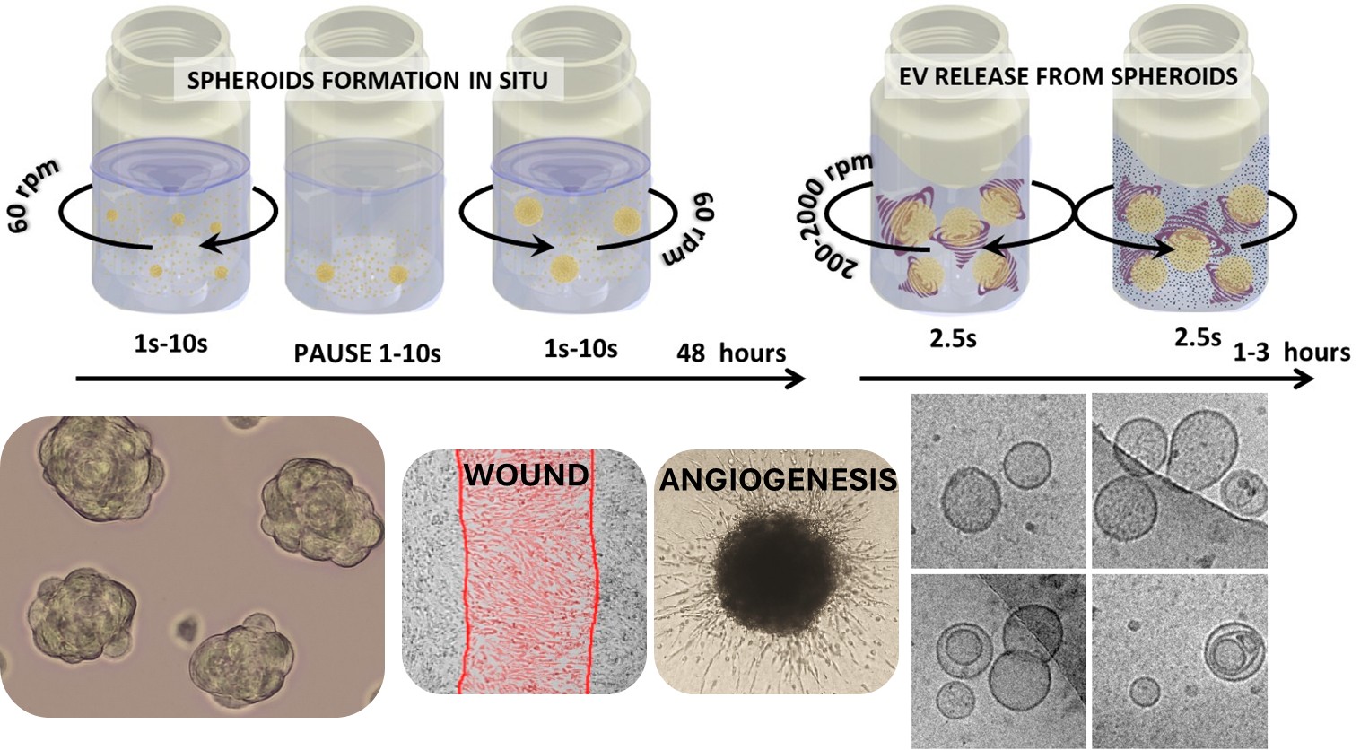
Hydrodynamic shear stress stimulation for the mass production of extracellular vesicles: the approach is based on a uniquely designed baffled tube undergoing alternating rotation, with validation on hMSC spheroids using a first laboratory-scale prototype.